


説明
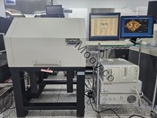
D3100S-1 Atomic Force Microscope (AFM) Key Features : . Scans samples up to 8 inch . Little or no sample preparation for increased productivity . Rigid ,low vibration construction for superior image quality . Integrated top-view color video optics with 1.5 um resolution and zoom . Easily changes among all AFM/STM scanning modes/techniques without tools . Automated stepping for scanning multiple areas unattended . Trakscan laser tracking system improves image and measurement quality . Laser spot alignment window for easy setup . Superior resolution and linearity in all three dimensions *. Fully refurbished. *. Installed in Clean-room. *. Can demonstrate any time.構成
- Dimension SPM Head - X-Y imaging area approx. 90um square - Z range approx. 6 um - Lateral accuracy typically within 1%, maximum 2% - Standard 150mm vacuum chuck for 100mm,125mm,150mm wafers - Optional 200mm vacuum chuck for 150mm and 200mm wafers - Inspectable Area 125x100mm ; allows coverage of one-half of 150mm wafer without manual sample rotation Full wafer with manual rotation - Nanoscope Dimension 3100 Controller - Nanoscope IIIa Scanning Probe Microscope Controller - System Computer - TMC Micro-G Isolation TableOEMモデルの説明
The Dimension 3100 Scanning Probe Microscope (SPM) is a high-resolution imaging device that produces three-dimensional images of a sample surface. The microscope works by scanning a sharp tip, mounted on a flexible cantilever, over the sample surface. The cantilever is attached to one end of a cylindrical piezoelectric tube, which is mounted near the top of the microscope. By applying voltages to the X, Y, and Z electrodes on the piezoelectric tube, the tube can be deflected horizontally and vertically to produce a precise raster scan over the sample surface. A stepper motor translates a slide with the sample attached, while a separate motor drive controls the height of the microscope and tip relative to the sample surface. This allows for accurate and detailed imaging of the sample surface.ドキュメント
ドキュメントなし

カテゴリ
Microscope
最終検証: 60日以上前
主なアイテムの詳細
状態:
Used
稼働ステータス:
不明
製品ID:
102010
ウェーハサイズ:
8"/200mm
ヴィンテージ:
不明
Logistics Support
Available
Transaction Insured by Moov
Available
Refurbishment Services
Available
同様のリスト
すべて表示


VEECO / DIGITAL INSTRUMENTS
DIMENSION 3100
カテゴリ
Microscope
最終検証: 60日以上前


主なアイテムの詳細
状態:
Used
稼働ステータス:
不明
製品ID:
102010
ウェーハサイズ:
8"/200mm
ヴィンテージ:
不明
Logistics Support
Available
Transaction Insured by Moov
Available
Refurbishment Services
Available
説明
D3100S-1 Atomic Force Microscope (AFM) Key Features : . Scans samples up to 8 inch . Little or no sample preparation for increased productivity . Rigid ,low vibration construction for superior image quality . Integrated top-view color video optics with 1.5 um resolution and zoom . Easily changes among all AFM/STM scanning modes/techniques without tools . Automated stepping for scanning multiple areas unattended . Trakscan laser tracking system improves image and measurement quality . Laser spot alignment window for easy setup . Superior resolution and linearity in all three dimensions *. Fully refurbished. *. Installed in Clean-room. *. Can demonstrate any time.構成
- Dimension SPM Head - X-Y imaging area approx. 90um square - Z range approx. 6 um - Lateral accuracy typically within 1%, maximum 2% - Standard 150mm vacuum chuck for 100mm,125mm,150mm wafers - Optional 200mm vacuum chuck for 150mm and 200mm wafers - Inspectable Area 125x100mm ; allows coverage of one-half of 150mm wafer without manual sample rotation Full wafer with manual rotation - Nanoscope Dimension 3100 Controller - Nanoscope IIIa Scanning Probe Microscope Controller - System Computer - TMC Micro-G Isolation TableOEMモデルの説明
The Dimension 3100 Scanning Probe Microscope (SPM) is a high-resolution imaging device that produces three-dimensional images of a sample surface. The microscope works by scanning a sharp tip, mounted on a flexible cantilever, over the sample surface. The cantilever is attached to one end of a cylindrical piezoelectric tube, which is mounted near the top of the microscope. By applying voltages to the X, Y, and Z electrodes on the piezoelectric tube, the tube can be deflected horizontally and vertically to produce a precise raster scan over the sample surface. A stepper motor translates a slide with the sample attached, while a separate motor drive controls the height of the microscope and tip relative to the sample surface. This allows for accurate and detailed imaging of the sample surface.ドキュメント
ドキュメントなし